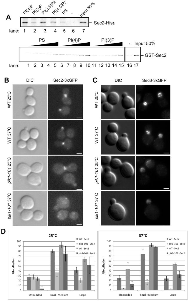
Figure 1.
(A) Sec2p directly binds to Phosphoinsitides. (top panel) Sec2p-His6 (120 nM) purified from E. Coli was incubated by itself (lane 6) or with 0.5 mM liposomes containing 50/30/15 mol% of PC/PE/PS and 5 mol% of the indicated phosphoinositides (lanes 1–4), or 45/30/25 mol% of PC/PE/PS (lane 5). Liposomes were precipitated by centrifugation at 100,000 × g and bound proteins were detected by Coomassie blue staining. (bottom panel) GST-Sec2p purified from E. Coli was incubated with 0.05, 0.1, 0.25, 0.375, and 0.5 mM of liposomes containing 10 mol% of PS, PI4P, or PI3P. Liposomes were precipitated by centrifugation at 100,000 x g and bound proteins were detected with anti-Sec2p antibody. (B) Sec2p is mislocalized in pik1-101 cells. Localization of Sec2-3xGFP (B) and Sec6-3xGFP (C) was examined in wildtype (WT) and pik1-101 cells. Cells were grown overnight at 25°C in a synthetic medium containing 2% glucose and then shifted to 37°C for 60 minutes. Cells were immediately fixed and examined as described in Experimental procedures. Bars are 2 μm. (D) Cells were classified into three categories (unbudded, small/medium buds, or large buds). A total of approximately 200 cells were counted for each experiment. Values indicate the percentage of cells showing bud or mother-daughter neck localization of Sec2-3xGFP or Sec6-3xGFP. Mean and S.D. of three different experiments are shown.

