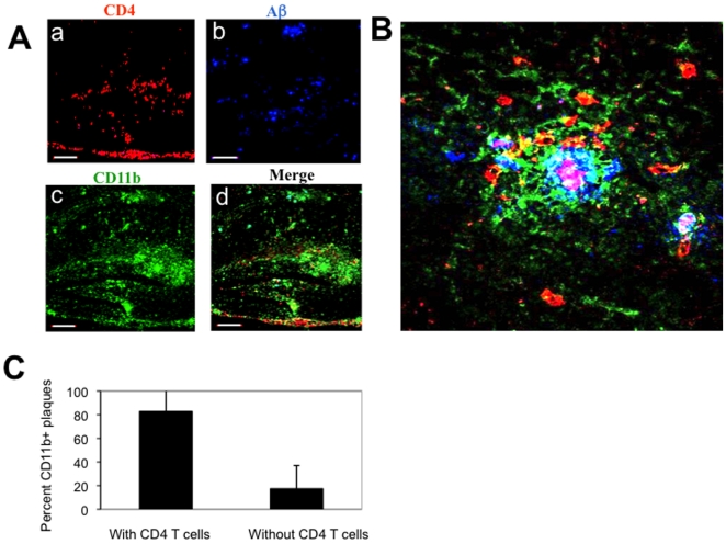
Figure 1. Aβ immunization results in trafficking of T cells to sites of Aβ plaques in the brain parenchyma.
APP/IFN-γ Tg mice aged 9 months were immunized with Aβ and killed 19 days later. Brain sections were immunolabeled for Aβ plaques co-localized with lymphocyte subpopulations and activated microglia. (A) Brain sections derived from a representative Aβ-immunized APP/IFN-γ Tg mouse (n = 11) were immunolabeled with anti-CD4 (a, red), anti-Aβ (b, blue), and anti-CD11b (c, green) antibodies. The merged panel is shown in d. Bar represents 200 µm. (B) Three-dimensional representation of Z-stalk images taken from a representative compact Aβ plaque co-localized with CD11b+ microglia (green) and CD4 T cells (red) in the hippocampus. Bar represents 20 µm. (C) Co-localization analysis of CD4 T cells and CD11b-labeled plaques. Using Volocity 3D image analysis software (Improvision, Waltham, MA, USA), we set an intensity threshold to mark only those areas with CD11bhigh microglia, representing Aβ plaques. Columns represent percent CD11b-labeled plaques with and without co-localized CD4 T cells in each of the analyzed sections [n = 3 (3 brain sections per mouse); means ± SD; P<0.001, Student's t test].

