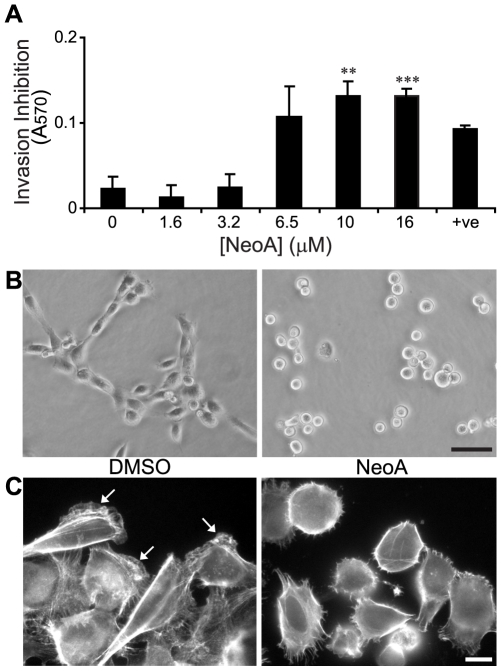
Figure 1. NeoA prevents tumor cell elongation and invasion into Matrigel.
(A) MDA-MB-231 cells were treated with NeoA (or 5 µM dihydromotuporamine C [10] as a positive control, +ve). Live cells that failed to invade were recovered and quantified through an MTT assay. Shown are averages of triplicates ± SD. ** P<0.005, *** P<0.0005 compared to 0 µM as determined by two-tailed Student's t-test. (B) MDA-MB-231 cells were plated on the reconstituted basement membrane substratum Matrigel in the presence of DMSO vehicle alone or 6.5 µM NeoA and morphology was assessed by live phase contrast microscopy after 2.5 h. Scale bar, 50 µm. (C) NeoA treatment results in loss of polarity and decreased actin ruffling. f-actin staining of cells treated with either DMSO or 3.2 µM NeoA for 90 min. Note that f-actin-rich leading lamellae (white arrows in DMSO-treated cells) are greatly reduced in the NeoA-treated cells. Scale bar, 10 µm.

