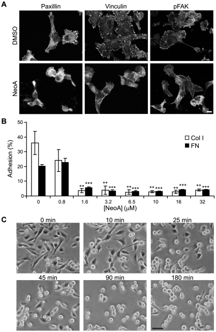
Figure 2. NeoA inhibits cellular adhesion and causes the disassembly of focal adhesions in MDA-MB-231 cells.
(A) Cells treated for 1 h with DMSO or 6.5 µM NeoA were fluorescently stained for the focal adhesion proteins paxillin, vinculin, and phosphorylated focal adhesion kinase (pFAK). Note that all proteins were no longer localized at discrete attachment sites (i.e. focal adhesions) in NeoA-treated cells. Scale bar, 10 µm. (B) Adhesion to fibronectin (FN) and collagen type I (Col I) is significantly decreased in the presence of NeoA. Shown are averages of triplicates ± SD. ** P<0.005, *** P<0.0005 compared to 0 µM as determined by two-tailed Student's t-test. (C) Cells pre-attached on fibronectin begin to lose adherence between 10 to 25 min after the start of treatment with 6.5 µM NeoA. Scale bar, 50 µm.

