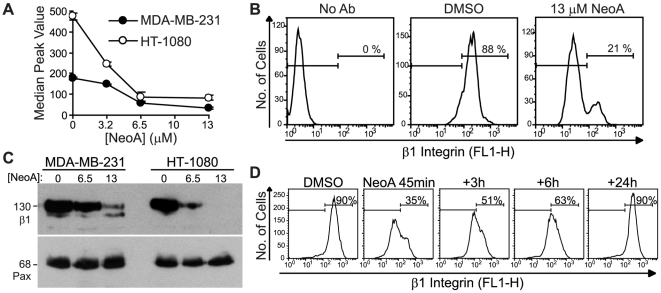
Figure 4. NeoA reversibly reduces levels of β1 integrin on the cell surface.
(A–B) MDA-MB-231 and HT-1080 cells treated in suspension with NeoA for 45 min were stained with a FITC-labeled antibody for total β1 integrin and surface levels were evaluated by flow cytometry. (A) Graphical representation of the shift in the median peak values (averaged from duplicates within one experiment ± SD) of β1 integrin after treatment with the NeoA. (B) NeoA lowers the percentage of MDA-MB-231 cells expressing β1 integrins on their surface. (C) Immunoblots of whole cell lysates from cells treated with 0, 6.5, or 13 µM of NeoA for 45 min. A representative blot from three independent experiments was probed for β1 integrin (β1) and paxillin (Pax). (D) Decrease in β1 integrin is reversible. MDA-MB-231 cells treated for 45 min with 13 µM NeoA were washed and allowed to recover for the indicated times. Surface levels of β1 integrin were measured by flow cytometry. All data shown are representative from at least three independent experiments.

