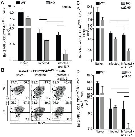
Figure 2. Simultaneous deficiency of IL-7 and IL-15 results in poor development of IL-7RαhiBcl-2+CD8+ T cells.
WT, KO, anti IL-7 treated WT and anti IL-7 treated KO mice were sacrificed at day 14 p.i. A–D, Splenocytes were surface stained for CD8β, CD44 and CD127, followed by intracellular staining for Bcl-2. A, Bcl-2 MFI of CD8+CD44int/hi T cells is shown as bar graph. B–D, Data of activated CD8+ T cell subsets are presented as Bcl-2 vs. CD127 density-contour plot (B) or as bar graphs of Bcl-2 MFI of CD8+CD44int/hiCD127hi T cells (C) or CD8+CD44int/hiCD127hiBcl-2+ (D). The experiment was performed at least twice with similar results. The data is representative of one experiment with 4 mice per group.

