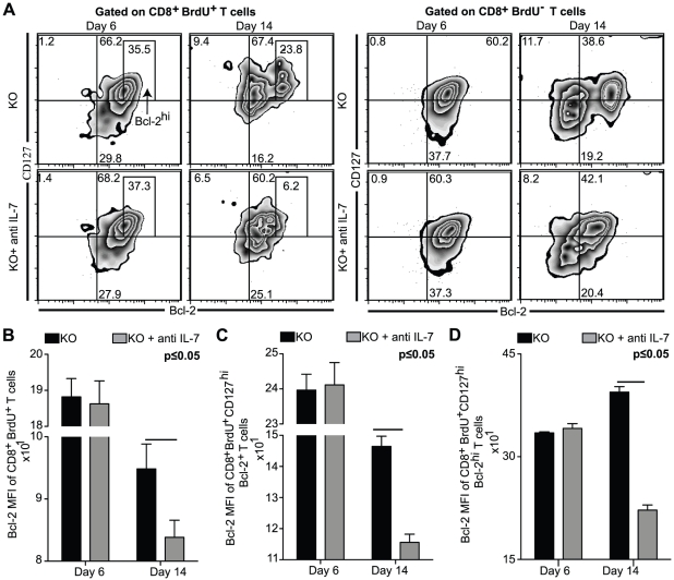
Figure 5. Bcl-2 and IL-7Rα are downregulated in BrdU+CD8+ T cells from IL-7 depleted KO mice.
Splenocytes from BrdU injected antibody or saline treated KO mice were harvested at day 6 and day 14 p.i. The cells were stained for CD8β, CD127, Bcl-2 and BrdU as described in Materials and Methods . A, The Bcl-2 vs. CD127 density-contour plots depicted on the top panel are gated on CD8+Brdu+ T cells (top left panel) and CD8+BrdU− T cells (top right panel). B–D, Bcl-2 MFI of CD8+BrdU+ T cells (B), CD8+BrdU+CD127hiBcl2+ T cells (C) and CD8+BrdU+CD127hiBcl2hi T cells (D) are presented as bar graphs in the lower panel. The experiment was performed twice with similar results. The data is representative of one of two similar experiments with 3–4 mice per group.

