
Fig. 3.
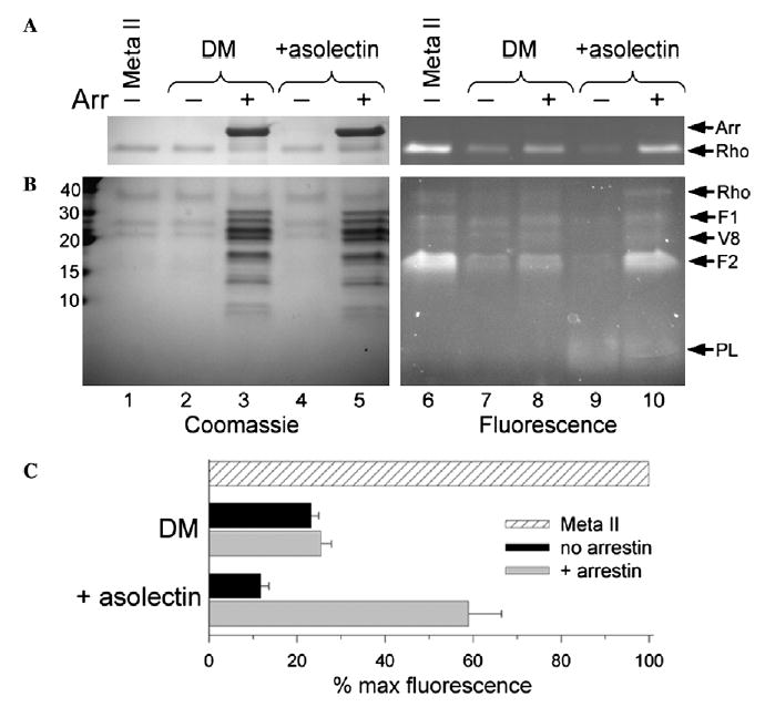
Arrestin traps retinal as a Schiff-base adduct on F2 of Rho*-P. (A) Reducing the retinal Schiff-base with NaBH4 produces a fluorescent Rho*-P that indicates the amount of retinal covalently attached to opsin. Briefly, Rho-P solubilized in 0.02% DM or 0.02% DM and 0.02% asolectin, without or with a twofold excess of arrestin, was photoactivated, allowed to decay at 20 °C in the dark, reduced after 120 min, and then subjected to SDS–PAGE. The total amount of Schiff-base linked retinal was assessed by the reduction of Meta II Rho immediately after bleach (lanes 1 and 6). (B) The same samples as described in (A) were subjected to V8 proteolysis. Molecular weight markers (kDa) are indicated on the left. In both (A) and (B), the Coomassiestain (left, lanes 1–5) and fluorescence (right, lanes 6–10) of each gel is shown, and arrows on the right identify the bands: arrestin (Arr), rhodopsin (Rho), Rho fragments resulting from V8 proteolysis (F1 and F2), the V8 protease (V8), and asolectin phospholipids (PL). (C) Plot of the average quantified fluorescence from four independent experiments as shown in (A). The fluorescence is expressed as a percent of the total “Meta II” fluorescence, and the error bars represent the standard error.