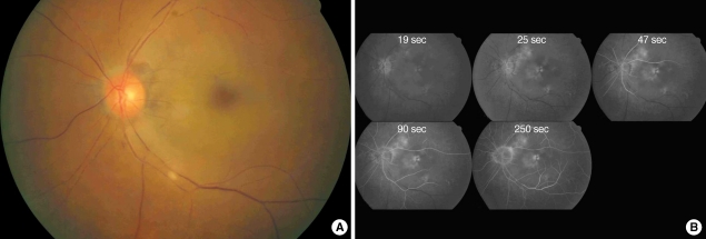
Fig. 1.
Fundus photography (A) and fluorescein angiography (B) before intra-arterial thrombolysis in case 1. There was white edematous retina with typical cherry-red spot and fragmentation of retinal vessels compatible with central retinal artery occlustion (A). Retinal arterial filling was markedly delayed with arterio-venous transit time of 5 min. The choroidal perfusion was normal (B).

