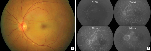
Fig. 3.
Fundus photography (A) and fluorescein angiography (B). One day after intra-arterial thrombolysis in case 1. Perfusion of retinal vessels were normalized and retinal color was restored to normal orange hue. However, there still remained macular edema (A). Retinal arterial and venous filling was normalized with arterio-venous transit time was 18 sec (B).

