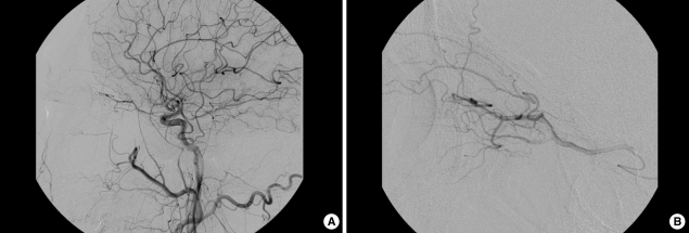
Fig. 5.
Common carotid angiogram in case 2 (A). There was no definite occlusion and remarkable lesion in ophthalmic artery and main trunk of intracerebral arteries. Selective angiogram of ophthalmic artery after intra-arterial thrombolysis (B). The muscular branches and ciliary arteries were well shown.

