
Figure 2.
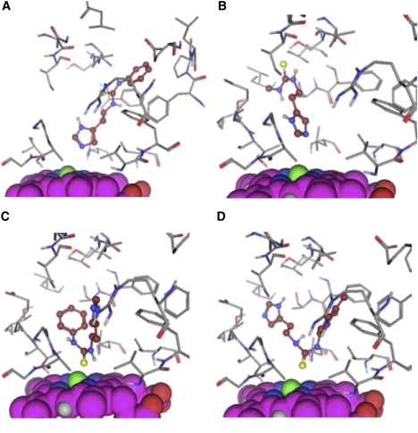
Examples of the four different starting conformations for the MD simulations. The heme is displayed in space-fill and is oriented in the same way in all figures. The ligand is displayed in ball-and-stick, with carbon atoms and the residues of the protein in sticks. (A) N_1 pose of compound TH6. (B) N_2 pose of compound TH1. (C) Pose S_1 of compound TH6. (D) Pose S_2 of compound TH7.