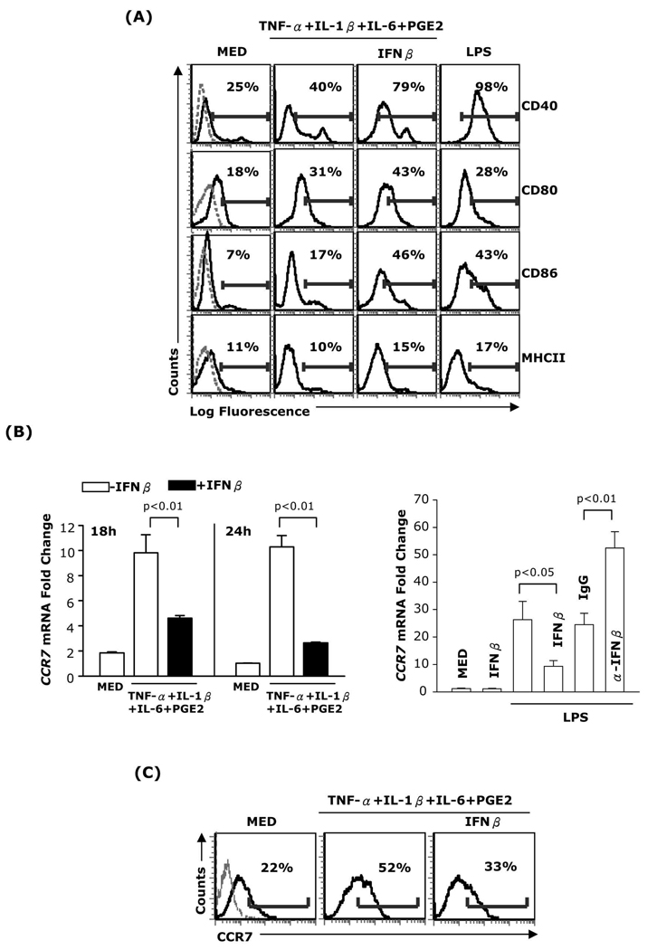
Fig. 1. Effects of IFNβ on DC maturation markers and CCR7 expression.
(A) DC were treated for 24h with TNF-α (20ng/ml), IL-1β (10ng/ml), IL-6 (10ng/ml), and PGE2 (10−6M) in the presence or absence of IFNβ (1,000 IU/ml), or LPS (1µg/ml). The expression of MHCII, CD40, CD80, and CD86 was analyzed by FACS. Dotted lines in the left panels represent isotype controls. (B, left panel) RNA was extracted from DC treated as above and subjected to real-time RT-PCR for CCR7. (B, right panel) DC were treated with LPS in the absence or presence of IFNβ, with or without neutralizing anti-IFNβ antibody (10µg/ml) or control IgG (10µg/ml) for 24h. Neutralizing anti-IFNβ antibody and control IgG were added twice: 30min before and 12h after LPS. RNA was extracted and subjected to real-time RT-PCR for CCR7. (C) DC were treated with the cytokine cocktail in the presence or absence of IFNβ for 24h and surface CCR7 expression was analyzed by FACS. Data are representative of three independent experiments.

