
Fig. 1.
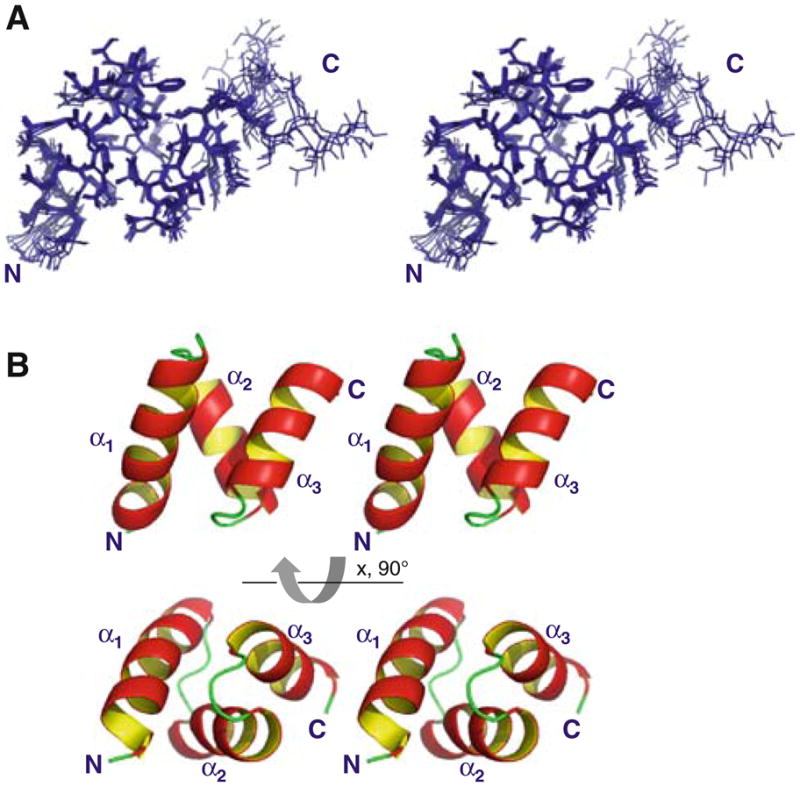
Solution structure of the UBA domain of CIP75. A. Stereo-view of the 10 lowest energy structures which have been superposed according to the backbone atoms (region M549-S593). B. Stereoview of the ribbon diagram of the lowest energy CIP75 UBA domain structure. The three α-helices (α1, α2, and α3) and the amino (N) and carboxyl (C) terminal domains have been labeled