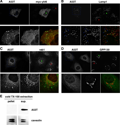
Figure 5.
Subcellular distributions of α-synuclein A53T, coexpressed myc-ykt6, and the unperturbed endogenous cellular transport machinery. (A) Comparison between α-synuclein A53T and myc-ykt6 staining patterns. (B) Comparison of α-synuclein A53T and endogenous LAMP-1. Blue arrowheads highlight a few of the α-synuclein-positive particles that are positive for LAMP-1. (C) Comparison of α-synuclein A53T and endogenous rab1. (D) Comparison of α-synuclein A53T and endogenous GPP130. In each block shown, green asterisks mark the nucleus of an α-synuclein A53T-expressing cell and red asterisks mark the nucleus of an untransfected cell for comparison. Shown are individual focal planes of deconvolved widefield images. (E) A53T-transfected NRK cells were extracted with cold 1% Triton X-100 and centrifuged at 10,000 × g for 15 min to generate a supernatant and pellet. Equal proportions of these fractions were analyzed by SDS-PAGE and immunoblotting for α-synuclein and caveolin, a protein known to be in detergent-resistant membrane domains.

