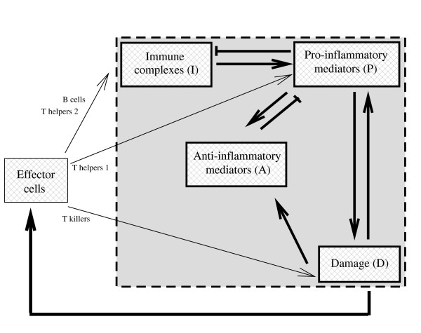
Figure 3.
Network of interactions that mediate renal damage in lupus nephritis. Naive T cells (not shown) are activated by the self-antigen presenting cells (APCs), and release cytokines and various chemical signals that stimulate the activity of other immune cells, such as natural killer cells, helper T cells, B cells and macrophages. Each of these activation pathways can lead to tissue destruction. Frequently, helper T cells can cause local inflammation and tissue damage by recruiting macrophages via cytokines and chemokines. Tissue damage can also occur directly via the activity of cytotoxic natural killer cells. However, extensive tissue damage is due to auto-antibodies, produced by the B cells. These auto-antibodies form immune complexes with self-antigen, either by binding directly to cell surface antigens, or by forming immune complexes in the circulation that deposit in the kidney. Immune complexes activate the complement system (not shown), which recruits and activates effector leukocytes (e.g. neutrophils, macrophages). These pro-inflammatory activated leukocytes produce toxic products that damage tissue. Concurrent activation of anti-inflammatory cells and production of anti-inflammatory mediators counterbalance the action of pro-inflammatory mediators. The flare process undergoes positive feedback because debris from apoptotic and damaged cells further stimulates the autoimmune response. As the flare is treated, activated effector cells are reduced, the production of auto-antibodies is disrupted, the deposition of immune complexes decreases, and tissue that is not permanently scarred undergoes repair or regeneration. Our mathematical model, Eqs (1)-(4), builds on the gray box interactions and follows the evolution in time of four variables: immune complexes (I), pro-inflammatory mediators (P), damaged tissue (D), and anti-inflammatory mediators (A).

