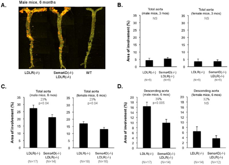
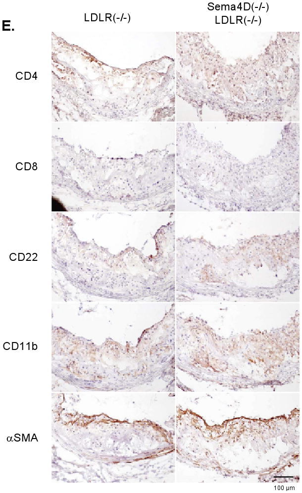
Figure 4. Loss of sema4D expression reduces atherosclerotic lesion size.


(A) Aortas after 6 months on the high fat diet showing lipid-rich deposits. (B,C,D) Analysis of lesion size. (E) Sections from the aortic root. Markers: CD4 and CD8, T-cells; CD22, B-cells; CD11b, monocytes, macrophage and neutrophils; smooth muscle actin (αSMA), smooth muscle cells and fibrotic caps. No differences were observed.
