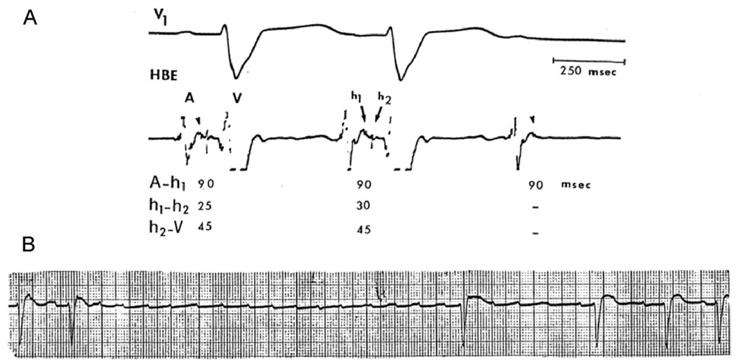
Figure 1.
Electrocardiograms (ECGs) from a 65-year-old woman with a history of recurrent syncopal episodes showed normal sinus rhythm, complete left BBB pattern, and periods of Mobitz type II AV block. A: Electrophysiological study (EPS) showed intra-His bundle block with split His bundle potential (h1, h2) and a 5-ms increment of the h1, h2 interval before block. Intra-His bundle block was initially unrecognized because the h1 deflection was overlooked. B: Rhythm strip 1 day later shows atrial tachycardia (rate 150–170 bpm) leading to TD-PAVB with 5.6-second ventricular asystole. AV conduction improved when the tachycardia slowed down (probably with a period of 2:1 AV block). Note the different time scale of the EPS recording.

