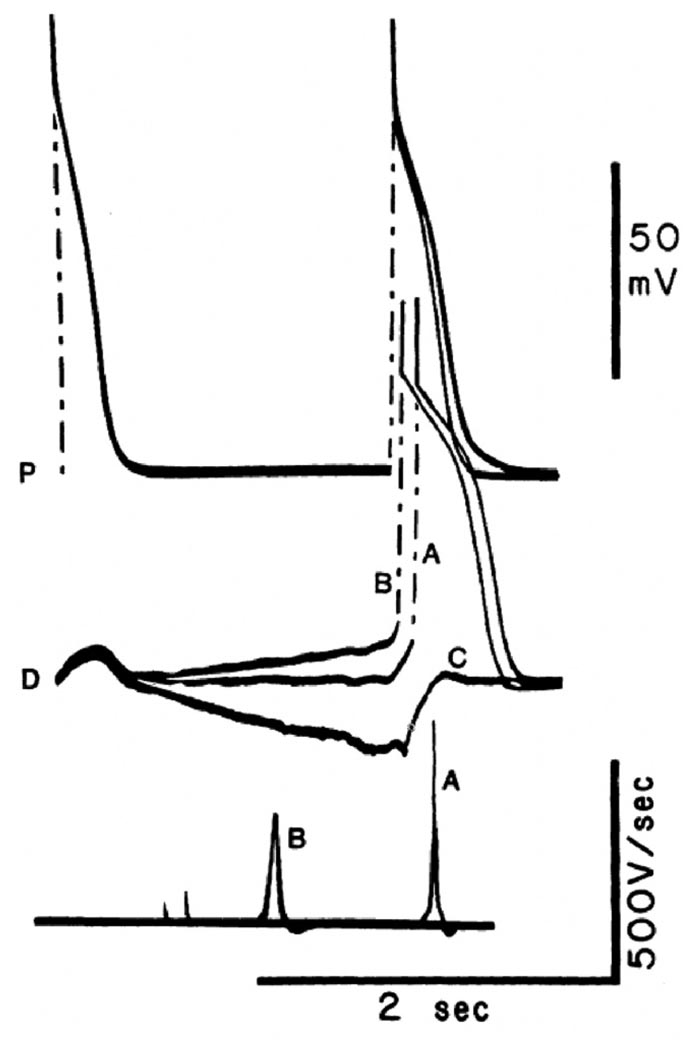
Figure 6.
Influence of diastolic depolarization versus hyperpolarization on conduction in a heterogeneous Purkinje fiber bundle placed in a three compartment tissue bath. Sucrose superfusion of the middle segment. The two outer fiber segments were superfused with normal Tyrode’s solution, stimulation was applied to the proximal segment (P), and activity was recorded from P and from the distal segment (D). Three superimposed recordings are shown. Top traces, P; middle traces, D; bottom traces dV/dt of D. In all sweeps, the initial action potential is the last of a series of 10 evoked in P at BCL = 500 ms. At this rate, complete PD block occurred, and only subthreshold depolarizations were apparent in the distal segment. A postmature action potential initiated in P after 1850 ms activated the distal fiber after 141 ms (discharge A). This long delay occurred despite the large amplitude (89 mV) and dV/dt max (475 V/s) of the distal action potential. In the next sweep, application of a slow depolarizing current ramp to D increased its slope of phase 4 depolarization. When, at the end of the ramp, the proximal segment was stimulated, propagation was again successful, but at a much reduced P-D conduction time (59 ms). Rapid propagation occurred even though the amplitude and dV/dt of phase 0 were significantly decreased in the distal segment, as a result of a reduction of the takeoff potential. In contrast, when the slope of phase 4 was inverted by the application of a hyperpolarizing ramp, complete block was induced (C), and only the electrotonic image of the proximal action potential was apparent in the distal trace. (Reproduced with permission of the American Heart Association from Jalife J, et al, Circulation 1983;67:912–922).

