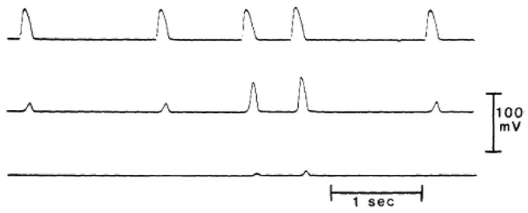
Figure 7.
PD conduction block in an isolated false tendon (dog) exposed to 20 mM KCL Tyrode’s solution. Top trace obtained from cell close to stimulation site; middle trace, from center of the bundle; and bottom trace, recorded more distally. At BCL = 1500 ms all impulses were blocked before they reached the middle segment. Successful activation of that segment was obtained when the stimulus interval was reduced to 500 ms. However, block persisted at the distal segment (reproduced with permission of the American Heart Association from Jalife J, et al, Circulation 1983;67:912–922).

