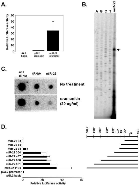
Figure 1. Identification of miR-22 promoter.
A. A 1155 bp fragment of miR-22 predicted promoter from −1100 to +55 was cloned upstream to the luciferase gene in the promoter-less pGL2 plasmid and transfected into HEK 293T together with RSV-renilla that served as internal control for transfection efficiency. pGL2-basic and pGL2-SV40-promoter were also transfected and served as negative and positive controls respectively. 24 hours later the relative luciferase activities were determined. The results represent the mean +/− SD of 7 independent transfection experiments. B. Determination of miR-22 TSS. The luciferase reporter gene under the control of miR-22 promoter was transfected into HEK293T cells and 24 hours after transfection RNA was extracted and analyzed by primer extension using a primer complementary to the 5′-end of the luciferase gene. The labeled miR-22 derived cDNA was run on a denaturing 8% urea-polyacrylamid gel alongside a sequencing reaction marked by A, G, C and T. C. Analysis of sensitivity of endogenous miR-22 transcription to α-amanitin. Nuclei were isolated from HEK293T cells and subjected to a run-on assay in the absence or presence of 20 µg/ml α-amanitin using 32P-labeled UTP. Labeled RNAs were isolated and then hybridized to membranes that were dot-blotted with RNA pol I gene, 45s rRNA; RNA pol III gene tRNAY and miR-22 transcripts as indicated. D. Analysis of miR-22 promoter organization. The miR-22 promoter was dissected from the 5′ as shown schematically and luciferase activity of the different constructs was determined as described in A. The results represent the mean +/− SD of 4–6 independent transfection experiments.

