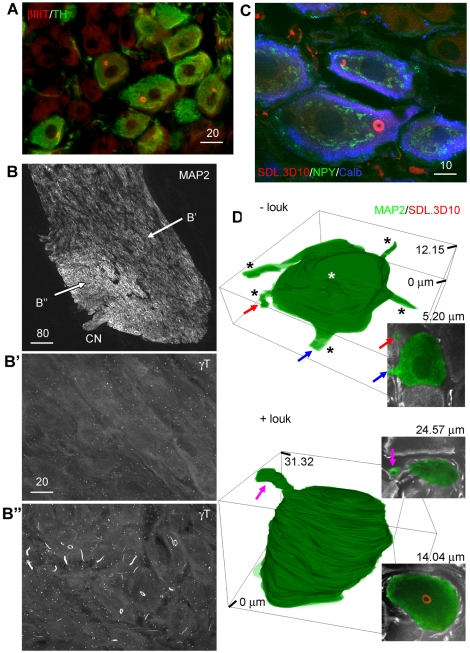
Figure 2. Distribution of the loukoumasome among neurons.
A, Loukoumasome-containing neurons are TH-positive (pelvic ganglion, standard epifluorescence). B, In the stellate ganglion, loukoumasome-containing neurons are larger, more densely packed, and situated near the exit point of the cardiac nerve (CN). MAP2: microtubule-associated protein 2. B′ and B″ indicate the regions enlarged in the lower panels (confocal stacks, maximum projections). C, Loukoumasomes occur exclusively in a subset of neurons expressing both neuropeptide Y (NPY) and calbindin-d28k (Calb) (stellate ganglion, single confocal slice). D, Neurons with loukoumasomes (+louk) in the stellate ganglion have fewer processes than those without (-louk). Insets: confocal slices at the depths indicated. Coloured arrows indicate the same processes in confocal slices and 3D reconstructions. See also Videos S1 and S2. Scale bar units: µm.

