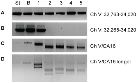
Figure 6. Marking of the breakpoint and detection of de novo telomere formation by PCR in sgs1Δ exo1Δ CanR HphS cells.
From the BIR assay. (A) PCR analysis of a starting strain prior to DSB induction (ST), CanS HphS colony that has repaired by BIR (B), and five CanR HPHS colonies (1–5) with primers that amplify sequences (Ch V 32,763–34,020) approximately 750 bp proximal to the break. (B) PCR with primers that amplify sequences (Ch V 32,265–34,020) approximately 250 bp proximal to the break. (C) PCR with a Ch V-specific primer that amplifies all colonies indicated and primer CA16, a telomere-specific primer. (D) PCR product from 6C ran longer an agarose gel to better display the laddered PCR product indicative of de novo telomere formation in samples 1–5.

