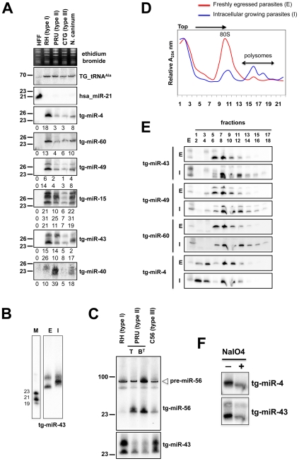
Figure 4. Expression patterns and characteristics of representative microRNAs in Toxoplasma.
(A) Expression profile analyses of six parasite microRNA candidates by small-RNA Northern hybridization in three canonical strains of Toxoplasma and its close relative, Neospora caninum. To control for loading, blots were stripped and rehybridized with the indicated miRNA and the control probes (tRNAAla and hsa_miR-21). Ethidium bromide staining of rRNA was used as loading control. Hybridization signals were quantified with the ImageJ software, normalized to the amount of Toxoplasma tRNAAla signal present in each sample, and shown as digit below each Northern blot. For miR-15, -43 and -49, each individual signal was separately quantified. The number next to each panel represents the position of RNA markers. Tg, Toxoplasma gondii; hsa, Homo sapiens. (B) Total RNA from freshly egressed (E) and intracellular growing parasites (I) were analyzed by Northern hybridization with complementary probes for Tg-miR-43. RNA markers (left lane) are 19, 21 and 23 nucleotides. (C) Total RNA from types I, II (tachyzoite and pH-converted bradyzoite) and III were analyzed by Northern hybridization with complementary probes for Tg-miR-43 and -miR-56. Pre-miR indicates the foldback RNA precursor form; Tg-miR refers to the mature, predominant form. The number next to each panel represents the position of RNA markers. (D) Total RNA from freshly egressed (E, in red) and intracellular growing parasites (I, in blue) were subjected to sedimentation on 5%–45% sucrose gradients in the presence of cycloheximide (to preserve the association of translating ribosomes with mRNAs). Measurements of the total RNA in the gradient fractions reveal signature patterns of peaks corresponding to messenger-ribonucleoprotein complexes (mRNP), followed by the 80S ribosomal fractions and the polyribosomal fraction. As can be appreciated from the A254 profiles, the rapid proliferation of tachyzoites inside of the host cell (in blue) is diagnosed by the high level of translating polyribosomes. (E) A proportion of Tg-miRNAs cosediment with polyribosomes. Total RNA was extracted from each fraction and analyzed by Northern blots with complementary probes for 6 Tg-miRNAs [miR-43, -49, and 60 (shown) and mir-4, -15, and 40, (not shown)]. All of the Tg-miRNAs tested are predominantly localized to the mRNP with a clear shift to polyribosome fractions in intracellular growing parasites. (F) The 3′ end of Tg-miR-4 and -43 are not modified. Gel-fractionated small RNAs were examined by b-elimination assay (see methods).

