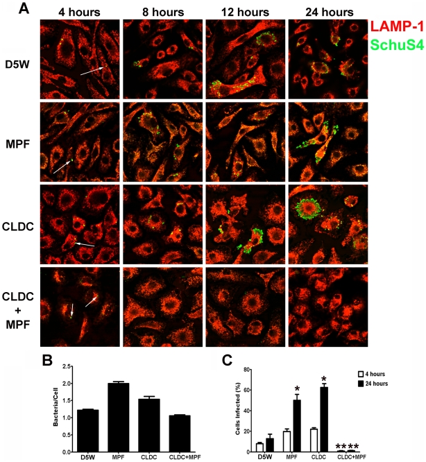
Figure 1. CLDC+MPF mediated control of F. tularensis replication in mouse macrophages.
Mouse macrophages were treated with D5W (untreated), MPF, CLDC, or CLDC+MPF 18 h prior to infection. Four, 8, 12 and 24 h after infection phagocytosis and intracellular replication of F. tularensis was monitored by microscopy. Exposure to CLDC, MPF or CLDC+MPF did not impact the number of cells infected (A and C) nor the number of bacteria entering each cell (B). MPF and CLDC treatment alone significantly increased the number SchuS4 infected cells 24 h after infection compared to untreated controls (* = p<0.01) (A and C). In contrast, CLDC+MPF treated cultures had fewer infected cells within 8 h of infection through 24 h after infection (** = p<0.01) (A and C). White arrows indicate intracellular SchuS4. Data is representative of four experiments. Error bars represent SEM.

