
Fig. 1.
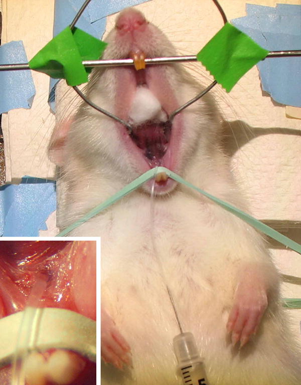
A cannula is inserted into the main (Wharton’s) excretory duct of the left submandibular gland of a female rat. A syringe carrying dispersed cells lies on the abdomen with the needle inserted into the distal end of the cannula. Insert: detail of the cannula inserted into Wharton’s duct.