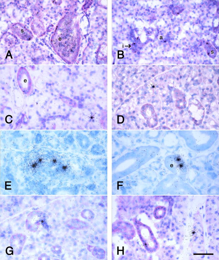
Fig. 3.
Autoradiographs of sections of female rat submandibular glands infusd with dispersed submandibular gland cells from male rats. The donor cells are identified by radiolabeled in situ hybridization by a Y-chromosome probe as in Figure 2E and F. The times post-infusion are 1 h (A, B), 1 day (C), 14 days (D, E, F) and 21 days (G, H). At 1 h, labeled cells pack the lumens of the excretory and striated ducts and many have lodged among the cells of both ducts and acini. Labeled cells have lodged in and have the cytodifferentiation of excretory ducts (e) in A, C, E, and H, striated ducts (s) in A, B, and E, intercalated ducts (i) in B, E, and G, and acini (a) in B, C, D, E, and H. A. B, C, D, G and H, basicfuchsin and toluidine blue stains; E, F, Giemsa stain. Scale bar = 70 μm for A and 50 μm for B through H.

