
Fig. 3.
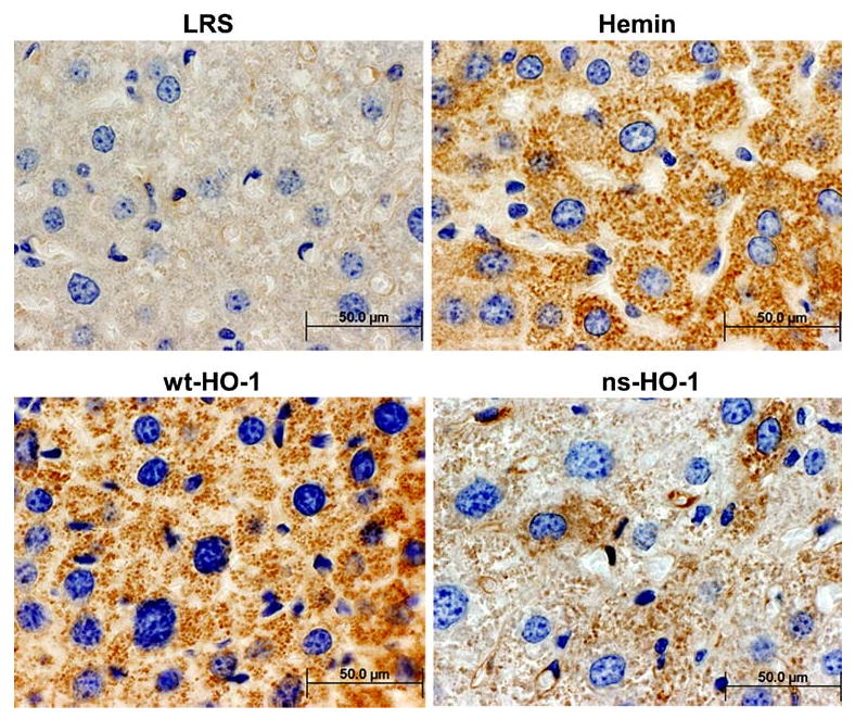
Immunohistochemistry demonstrates HO-1 overexpression in hepatocytes. Formalin-fixed paraffin embedded liver tissues were sectioned and stained with anti-HO-1. Sites of primary antibody binding were visualized with HRP-conjugated secondary IgG. HO-1/HRP immunoconjugates were detected with diaminobenzidine and H2O2. Nuclei were counter-stained with hematoxylin