Abstract
New benzophenone and flavonol galloyl glycosides were isolated from an 80% MeOH extract of Psidium guajava L. (Myrtaceae) together with five known quercetin glycosides. The structures of the novel glycosides were elucidated to be 2,4,6-trihydroxybenzophenone 4-O-(6″-O-galloyl)-β-d-glucopyranoside (1, guavinoside A), 2,4,6-trihydroxy-3,5-dimethylbenzophenone 4-O-(6″-O-galloyl)-β-d-glucopyranoside (2, guavinoside B), and quercetin 3-O-(5″-O-galloyl)-α-l-arabinofuranoside (3, guavinoside C) by NMR, MS, UV, and IR spectroscopies. Isolated phenolic glycosides showed significant inhibitory activities against histamine release from rat peritoneal mast cells, and nitric oxide production from a murine macrophage-like cell line, RAW 264.7.
Keywords: Psidium guajava L., Benzophenone galloyl glycoside, Flavonol galloyl glycoside
Introduction
Psidium guajava (Myrtaceae) is a small medicinal tree that is native to South America. All parts of this tree, including fruits, leaves, bark, and roots, have been used for treating stomachache and diarrhea in many countries. Previous studies of this plant have led to the isolation of tannins [1, 2], tannins and other phenolic compounds [3], flavonol glycosides [4, 5], triterpenoids [6], terpenoids [7], and carotenoids [8]. It is well known that an extract of the leaves of P. guajava improves symptoms of allergic disease, and we are interested in small molecules from the leaves of P. guajava, other than tannins, and their potential anti-inflammatory activities.
In this investigation, two new benzophenone galloyl glycosides, guavinosides A (1) and B (2), and a quercetin galloyl glycoside, guavinoside C (3), were isolated from the leaves of P. guajava together with known quercetin glycosides (4–8). These structures of the novel glycosides were established through detailed analysis of their spectroscopic data and chemical evidence, and their inhibitory activities against histamine release from rat mast cells and nitric oxide (NO) from RAW 264.7 were also determined.
Results and discussion
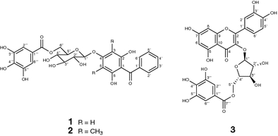
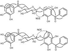
Guavinoside A (1) was obtained as a yellow powder. The positive ion mode FAB-MS of 1 showed a quasimolecular ion peak at m/z 545 [M + H]+, and the molecular formula was determined to be C26H24O13 based on its high-resolution (HR)-FAB-MS (found 545.8418, calcd. 545.8413 for C26H25O13). In the IR spectrum, the strong absorbance at 1,700 cm−1 indicated the presence of conjugated carbonyl groups in 1. Acid hydrolysis of 1 furnished d-glucose which was identical with HPLC analysis, using an optical rotation (OR) detector. In the 1H-NMR spectrum of 1, two aromatic signals at δ H 6.11 (2H, s) and 6.98, (2H, s) indicated the presence of two 1,3,4,5-tetrasubstituted phenyl groups, and the spin system of δ H 7.68 (2H, dd, J = 7.0, 1.0 Hz), 7.46 (2H, t, J = 7.0 Hz), and 7.57 (1H, t, J = 7.0 Hz) showed the presence of a phenyl group. Additionally, an anomeric proton signal at δ H 4.86 (1H, d, J = 7.6 Hz) indicated that the glucose residue was in the β-form. Correlations in the 1H–1H-COSY spectrum were observed from the anomeric proton to δ H 3.15 (1H, m), 3.20 (1H, m), 3.50 (1H, m), 3.30 (1H, m), and 4.39 (2H, br s) indicating the presence of β-glucose. The signal pattern in the aromatic region of the 13C-NMR spectrum indicated the presence of three aromatic rings. In addition, the 13C-NMR and DEPT spectra showed an anomeric carbon (δ C 100.5), four oxymethines (δ C 73.0, 76.1, 68.9, and 73.6), an oxymethylene (δ C 62.5), and two carbonyl carbons (δ C 165.7 and 195.3). All proton–carbon connectivities assigned by using HMQC experiments are summarized in Table 1. The HMBC correlations from 2″′, 6″′-H (δ H 6.98, 2H, s) to C-1″′ (δ C 119.2), C-3″′, 5″′ (δ C 145.4), C-4″′ (δ C 138.4), and a carbonyl carbon (δ C 165.7) revealed the presence of a galloyl moiety. A phenyl proton signal at δ H 7.68 (2H, dd, J = 7.0, 1.0 Hz, 2′, 6′-H) correlated with a carbonyl carbon signal at δ C 195.3, and an aromatic proton at δ H 6.11 (2H, s, 3, 5-H) correlated with C-4 (δ C 159.8) and the carbonyl carbon by weak 4 J correlation. Furthermore, the anomeric proton signal correlated with C-4. NOE enhancement was observed between the anomeric proton signal and the signal of 3, 5-H. These data suggested that the aglycone of 1 was 2,4,6-trihydroxybenzophenone, and a sugar was attached at C-4. The signal of 6′′-H, however, was downfield shifted at δ H 4.39 (2H, br s), and correlated with the galloyl carbonyl carbon signal at δ C 165.7 in the HMBC experiment. From these data, the structure of 1 was established to be 2,4,6-dihydroxybenzophenone 4-O-(6″-O-galloyl)-β-d-glucopyranoside (Figs. 1, 2).
Table 1.
13C- and 1H-NMR spectral data of guavinosides A (1) and B (2)
| 1 | 2 | |||
|---|---|---|---|---|
| δ C | δ H | δ C | δ H | |
| Aglycone | ||||
| 1 | 108.9 s | 113.0 s | ||
| 2, 6 | 157.3 s | 151.8 s | ||
| 3, 5 | 94.9 d | 6.11 (2H, s) | 110.8 s | |
| 4 | 159.8 s | 155.6 s | ||
| 1′ | 138.4 s | 138.7 s | ||
| 2′, 6′ | 128.7 d | 7.68 (2H, dd, J = 7.0, 1.0 Hz) | 128.6 d | 7.65 (2H, dd, J = 7.0, 1.0 Hz) |
| 3′, 5′ | 128.2 d | 7.46 (2H, t, J = 7.0 Hz) | 128.1 d | 7.45 (2H, t, J = 7.0 Hz) |
| 4′ | 132.5 d | 7.57 (1H, t, J = 7.0 Hz) | 132.3 d | 7.54 (1H, t, J = 7.0 Hz) |
| C=O | 195.3 s | 196.7s | ||
| 3, 5-CH3 | 9.9 q | 2.00 (6H, s) | ||
| Glucosyl | ||||
| 1″ | 100.5 d | 4.86 (1H, d, J = 7.6 Hz) | 104.2 d | 4.63 (1H, d, J = 8.5 Hz) |
| 2″ | 73.0 d | 3.15 (1H, m) | 74.2 d | 3.36 (1H, m) |
| 3″ | 76.1 d | 3.20 (1H, m) | 76.1 d | 3.30 (1H, m) |
| 4″ | 68.9 d | 3.50 (1H, m) | 69.3 d | 3.44 (1H, m) |
| 5″ | 73.6 d | 3.30 (1H, m) | 73.5 d | 3.42 (1H, m) |
| 6″ | 62.5 t | 4.39 (2H, br s) | 62.7 t | 4.23 (1H, d, J = 11.0, 3.0 Hz) |
| 4.32 (1H, d, J = 11.0 Hz) | ||||
| Galloyl | ||||
| 1″′ | 119.2 s | 119.6 s | ||
| 2″′, 6″′ | 108.6 d | 6.98 (2H, s) | 108.5 d | 6.96 (2H, s) |
| 3″′, 5″′ | 145.4 s | 145.3 s | ||
| 4″′ | 138.4 s | 138.3 s | ||
| C=O | 165.7 s | 165.6 s | ||
1H-NMR (600 MHz) and 13C-NMR (150 MHz) spectra were measured in DMSO-d 6 with TMS as internal standard
Fig. 1.

Structures of guavinosides A (1), B (2), and C (3)
Fig. 2.

Key HMBC and NOE correlations of guavinosides A (1) and B (2)
Guavinoside B (2) was obtained as a brownish solid. The negative ion mode FAB-MS of 2 showed quasimolecular ion peak at m/z 571 [M − H]−, and the molecular formula was determined to be C28H28O13 based on its HR-FAB-MS (found 571.1474, calcd. 571.1451 for C28H27O13). The molecular weight was 28 mass units (C2H4) greater than that of 1. The 1H- and 13C-NMR spectra of 2 were very similar to those of 1 except for absence of the aromatic methine signal [δ H 6.11 (s); δ C 94.9] in 1, and a new aryl methyl signal [δ H 2.00 (6H, s); δ C 9.9] and a quaternary carbon (δ C 110.8) were observed. In the 13C-NMR spectrum, the appearance of a high-field region shifted methyl signal suggested that the methyl is linked to a benzene ring in the ortho-position and attached via an oxygen atom [9]. HMBC correlations were observed from the methyl proton signal to δ C 110.8 (C-3, 5), 151.8 (C-2, 6), and 155.6 (C-4). NOE enhancement was also observed between the methyls and an anomeric proton at δ H 4.63. These data suggested that the aglycone of 2 was 2,4,6-trihydroxy-3,5-dimethylbenzophenone. Absolute configuration of the glucose moiety was determined to be d by using HPLC analysis with an OR detector. From the above data, the structure of 2 was identified to be 2,4,6-trihydroxy-3,5-dimethylbenzophenone 4-O-(6″-O-galloyl)-β-d-glucopyranoside.
Guavinoside C (3) was obtained as a yellow powder. Its HR-FAB-MS showed a quasimolecular ion peak at m/z 585.0868, corresponding to the molecular formula C27H22O15. The UV absorbances at 211, 265, and 355 nm were characteristic of flavonol. The IR spectrum indicated the presence of hydroxyl (3,400 cm−1), ester (1,710 cm−1), and conjugated carbonyl group (1,690 cm−1). In the 1H-NMR spectrum, meta-coupled signals at δ H 6.20 and δ H 6.41 and a hydrogen-bonded hydroxyl signal at δ H 12.62 indicated the presence of a 5,7-dihydroxy A ring system in flavonol. A spin system of three aromatic signals at δ H 7.46 (1H, d, J = 2.2 Hz), δ H 6.85 (1H, d, J = 8.8 Hz), and δ H 7.49 (1H, dd, J = 8.8, 2.2 Hz) indicated the presence of a 3′,4′-dihydroxy B ring system in flavonol. In the 13C-NMR spectrum, significant flavonol signals at δ C 157.3 (C-2), 133.0 (C-3), and 177.4 (C-4) were observed. In the HMBC experiment, the correlations from 2′-H to C-2, δ C 144.9 (C-3′), 148.3 (C-4′), and 121.3 (C-6′), from 6′-H to C-2, C-4′ and δ C 115.6 (C-2′), and from 5′-H to C-3′, C-4′, and δ C 120.8 (C-1′) were observed. From these data, the aglycone of 3 was determined to be quercetin. In addition, an aromatic methine (δ H 6.89, 2H, s) correlated with δ C 118.9 (C-1″′), 108.5 (C-2″′, 6″′), 145.3 (C-3″′, 5″′), 138.4 (C-4″′), and 165.4 (carbonyl), indicating the presence of a galloyl moiety the same as 1 and 2. Acid hydrolysis of 3 with 2 M HCl afforded (+)-l-arabinose that was identical by HPLC analysis using OR detector comparison to an authentic sample of l-arabinose. The small coupling constant of the anomeric proton (δ H 5.56, d, J = 1.4 Hz) indicated the presence of the α-form of arabinose. Correlations in the 1H–1H COSY spectrum were observed for a spin system from the anomeric signal to three oxymethine signals at δ H 4.18 (br s, 2″-H), 3.81 (m, 3″-H), and 3.74 (m, 4″-H), and methylene signals (δ H 4.11 and 4.02, 5″-H2) was observed. Furthermore, two hydroxy signals at δ H 5.72 (br s) and 5.48 (br s) both coupled with 2″-H and 3″-H, respectively. Additionally, the signals of 5″-H2 of 3 were shifted downfield compared with those of 4 (δ H 3.36 and 3.32), and correlated with a galloyl carbonyl carbon signal in the HMBC experiment. From these observations, the sugar moiety was determined to be l-α-arabinofuranose. Thus, the structure of 3 was determined to be quercetin 3-O-(5″-O-galloyl)-α-l-arabinofuranoside.
The structures of 4–8 were elucidated to be quercetin 3-O-α-l-arabinofuranoside (4), quercetin 3-O-α-l-arabinopyranoside (5), quercetin 3-O-β-d-xylopyranoside (6), quercetin 3-O-β-d-galactopyranoside (7), and quercetin 3-O-β-d-glucopyranoside (8) by comparison with spectroscopic data [10, 11] and chemical degradation methods.
Benzophenone glycosides have been isolated from many kind of plants, but this is first reported isolation of a dimethylbenzophene glycoside from a natural source. The substitution pattern is the well-known A ring of flavonoid; a possible biosynthesis pathway to the aglycone moiety of 2 would be methylation of the benzophenone skeleton.
Isolated compounds were evaluated for inhibitory activities against histamine release from rat peripheral mast cells [12] and nitric oxide (NO) production from a murine macrophage-like cell line, RAW264.7 cells [13]. Compounds 3–8 (at 100 μg/ml) inhibited histamine release from mast cells with inhibition ratios of 94.4, 21.9, 30.5, 23.9, 100, and 93.5%, respectively. But 1 and 2 did not show inhibitory activity against histamine release at this concentration. Compounds 3–8 (at 100 μg/ml) inhibited NO production by RAW 264.7 cells stimulated with lipopolysaccharide and interferon gamma with inhibition ratios of 50.0, 33.2, 32.4, 65.1, 55.3, and 52.1%, respectively. The isolated compounds therefore inhibited chemical mediators, such as histamine and NO, and increased IL-12 release from RAW 264.7 cells.
In conclusion, phenolic compounds isolated from P. guajava might be valuable candidates for treating various inflammatory diseases.
Experimental
General
The UV spectra were recorded on a Shimadzu model UV-160 spectrophotometer. IR spectra were recorded on a Horiba FT-210 diffraction infrared spectrometer. FAB-MS were obtained with a JEOL model JMS-AX505 HA spectrometer. 1H-NMR (600 MHz) and 13C-NMR (150 MHz) spectra were obtained on a Varian Inova™ 600 spectrometer and a JEOL Delta 600 spectrometer. NMR spectra were measured in DMSO-d 6 with TMS as internal standard. Optical rotation was measured with a Jasco DIP-370 polarimeter. The inhibitory activities against histamine release from rat mast cells and NO production were carried out as described in the literature method [14].
Plant material
Leaves of P. guajava were donated by OS Industrial Co. Ltd. (Tokyo, Japan).
Extraction and isolation
The dried leaves of P. guajava (5 kg) were extracted with 15 l of 80% MeOH at room temperature for 7 days. The solution was filtered and concentrated under reduced pressure to give a crude extract. The extract was dissolved in water and passed through a Diaion HP-20 column (Mitsubishi Kasei, Tokyo, Japan), and eluted stepwise with 50, 70, and 100% MeOH. The 70% MeOH eluate was dissolved in EtOH and passed through a Sephadex LH-20 column (Pharmacia, Uppsala, Sweden). The effluent was chromatographed on a Diaion CHP-20P column (Mitsubishi Kasei, Tokyo, Japan), and eluted with 50 and 70% MeOH. All fractions were monitored by TLC, and the fractions containing the same compound(s) (as evidenced by TLC) were combined to give four fractions. Fraction 3 (900 mg) was chromatographed on a Sephadex LH 20 column developed with CHCl3–MeOH (1:1) to give four fractions. Fraction 2 (770 mg) was further applied to a reversed-phase column (SSC ODS, Senshu Scientific Co. Ltd., Tokyo, Japan) eluted stepwise with 0–25% MeOH, and recrystallized from MeOH to give 1 (47 mg). Fraction 4 (124 mg) was purified by medium pressure liquid chromatography (Yamazen Baker-bond ODS Yamazen, Kyoto, Japan) column eluted with MeCN–MeOH–H2O (5:35:60) to give 2 (44 mg). Fraction 3 (130 mg) was purified by reversed-phase HPLC [column: Shiseido Capcell pak C18 UG120 (10-mm i.d. × 250 mm, Shiseido, Tokyo, Japan); mobile phase: MeCN–MeOH–H2O (5:30:65); flow rate: 3.0 ml/min; detection: UV at 254 nm] to give 3 (14 mg). The 100% MeOH eluate of Diaion HP-20 was dissolved in MeOH, and chromatographed on a Sephadex LH-20 column (2.5 × 100 cm) to give ten fractions. Fraction 3 (1.9 g) was chromatographed on a silica gel column developed with CHCl3–MeOH to give eight fractions. Fraction 6 (515 mg) was purified by reversed-phase HPLC [column: Shiseido Capcell pak C18 UG120 (10-mm i.d. × 250 mm); mobile phase: MeCN–H2O (18:82); flow rate: 3.0 ml/min.; detection: UV at 250 nm] to give 4 (20 mg), 5 (50 mg), 6 (62 mg), 7 (62 mg), and 8 (30 mg), respectively.
Guavinoside A (1) was obtained as a yellow powder.  (c = 0.1, MeOH); FAB-MS m/z 545 (M + H)+; HR-FAB-MS (found 545.8418, calcd for C26H25O13: 545.8413); UV
  nm (ε): 218 (26,800), 288 (16,200); IR
  cm−1: 3,390 (OH), 1,700 (ester). 1H- and 13C-NMR data, see Table 1.
Guavinoside B (2) was obtained as a yellow powder;  (c = 0.5, MeOH); FAB-MS m/z 571 (M − H)−; HR-FAB-MS (found 571.1474, calcd for C28H27O13: 571.1451); UV
  nm (ε): 218 (26,400), 283 (15,100); IR
  cm−1: 3,410 (OH), 1,710 (ester), 1,690 (C=O). 1H- and 13C-NMR data, see Table 1.
Guavinoside C (3) was obtained as a yellow powder;  (c = 1.0, MeOH); FAB-MS m/z 585 (M − H)−; HR-FAB-MS (found 585.0868, calcd for C27H21O15: 585.0880);
  nm (ε): 211 (32,000), 265 (16,200), 355 (10,000); IR
  cm−1: 3,400 (OH), 1,710 (ester), 1,690 (C=O). 1H- and 13C-NMR data, see Table 2.
1H-NMR (600 MHz) and 13C-NMR (150 MHz) spectra were measured in DMSO-d
6 with TMS as internal standard
Table 2.
13C- and 1H-NMR spectral data of guavinoside C (3)
| No. | δ C | δ H |
|---|---|---|
| 2 | 157.3 s | |
| 3 | 133.0 s | |
| 4 | 177.4 s | |
| 5 | 161.9 s | |
| 6 | 98.5 d | 6.20 (1H, d, J = 1.5 Hz) |
| 7 | 164.1 s | |
| 8 | 93.5 d | 6.41 (1H, d, J = 1.5 Hz) |
| 9 | 156.3 s | |
| 10 | 103.9 s | |
| 1′ | 120.8 s | |
| 2′ | 115.6 d | 7.46 (1H, d, J = 2.2 Hz) |
| 3′ | 144.9 s | |
| 4′ | 148.3 s | |
| 5′ | 115.4 d | 6.85 (1H, d, J = 8.8 Hz) |
| 6′ | 121.3 d | 7.49 (1H, d, J = 8.8, 2.2 Hz) |
| 5-OH | 12.62 (1H, s) | |
| Arabinosyl | ||
| 1″ | 107.7 d | 5.56 (1H, J = 1.4 Hz) |
| 2″ | 81.7 d | 4.18 (1H, br s) |
| 3″ | 76.9 d | 3.81 (1H, m) |
| 4″ | 81.9 d | 3.74 (1H, m) |
| 5″ | 62.5 t | 4.11 (1H, dd, J = 12.0, 3.3 Hz) |
| 4.02 (1H, dd, J = 12.0, 5.8 Hz) | ||
| 2″-OH | 5.72 (1H, br s) | |
| 3″-OH | 5.48 (1H, br s) | |
| Galloyl | ||
| 1″′ | 118.9 s | |
| 2″′, 6″′ | 108.5 d | 6.89 (2H, s) |
| 3″′, 5″′ | 145.3 s | |
| 4″′ | 138.4 s | |
| C=O | 165.4 s | |
Acid hydrolysis of 1–3
Compound 1, 2, or 3 (3.0 mg, each) was treated with 0.5 ml of 2 M HCl for 2 h at 110°C in a sealed tube. The reaction mixture was diluted with 1 ml of H2O, and extracted with an equal volume of EtOAc, and the water layer was evaporated to dryness. The residue was dissolved in H2O (200 μl) and subjected to HPLC analysis [column: Asahi pak NH2P-50, (4.6-mm i.d. × 250 mm, Showa Denko, Tokyo, Japan); mobile phase: MeCN–H2O (75:25); flow rate: 1.0 ml/min.; detection: OR detector (Shodex OR-2, Showa Denko, Tokyo, Japan) and RI (Shodex RI-72, Showa Denko, Tokyo, Japan), column temperature: 40°C]. Retention time and optical rotation of these samples (1, 2, and 3) were found to be 8.5 min (positive), 8.5 min (positive), and 6.0 min (positive), respectively. Retention time of standard samples of (+)-d-glucose, (+)-l-arabinose, (−)-d-arabinose, and (+)-d-xylose were found to be 8.5 min (positive), 6.0 min (positive), 6.0 min (negative), and 6.3 min (positive), respectively.
Acknowledgments
This work was supported in part by a Grant-in-Aids from the Ministry of Health and Welfare of Japan (No. 19980070), by a Special Research Grant-in Aid for the development of characteristic education and High-Tech Research Centers from the Ministry of Education, Science, Sports and Culture of Japan to Nihon University, and by the Japan–China Medical Association, and “Academic Frontier” Project for Private Universities matching fund subsidy from MEXT (Ministry of Education, Culture, Sports, Science and Technology) 2002–2007 and 2007–2010 of Japan.
Open Access
This article is distributed under the terms of the Creative Commons Attribution Noncommercial License which permits any noncommercial use, distribution, and reproduction in any medium, provided the original author(s) and source are credited.
References
- 1.Okuda T, Yoshida T, Hatano T, Yazaki K, Ashida M. Ellagitannins of the casuarinaceae, stachyurqaceae and myrtaceae. Phytochemistry. 1982;21:2871–2874. doi: 10.1016/0031-9422(80)85058-8. [DOI] [Google Scholar]
- 2.Okuda T, Yoshida T, Hatano T, Yazaki K, Ikegami Y, Shingu T. Guavins A, C and D, complex tannins from Psidium guajava. Chem Pharm Bull. 1987;35:443–446. [Google Scholar]
- 3.Okuda T, Hatano T, Yazaki K. Guavin B, an ellagitannin of novel type. Chem Pharm Bull. 1984;32:3787–3788. doi: 10.1248/cpb.32.3787. [DOI] [PubMed] [Google Scholar]
- 4.Lozoya X, Meckes M, Abou-zaid M, Tortoriello J, Nozzolillo C, Arnason JT. Quercetin glycosides in Psidium guajava L. leaves and determination of a spasmolytic principle. Arch Med Res. 1994;25:11–15. [PubMed] [Google Scholar]
- 5.Morales MM, Tortoriello J, Meckes M, Paz D, Lozoya X. Calcium-antagonist effect of quercetin and its relation with the spasmolytic propaties of Psidium gujava L. Arch Med Res. 1994;25:17–21. [PubMed] [Google Scholar]
- 6.Osman AM, Younes M, Garby EL, Sheta AE. Chemical examination of local plants. VI. Triterpenoids of the leaves of Psidium guajava. Phytochemistry. 1974;13:2015–2016. doi: 10.1016/0031-9422(74)85152-6. [DOI] [Google Scholar]
- 7.Smith RM, Siwatibau S. Aeaquiterpene hydrocarbons of Fijian guavas. Phytochemistry. 1975;14:2013–2015. doi: 10.1016/0031-9422(75)83115-3. [DOI] [Google Scholar]
- 8.Mercadantre AZ, Steck A, Pfander H. Carotenoids from Guava (Psidium guajava L.): isolation and structure elucidation. J Agric Food Chem. 1999;47:145–151. doi: 10.1021/jf980405r. [DOI] [PubMed] [Google Scholar]
- 9.Matsuzaki K, Tahara H, Inokoshi J, Tanaka H, Masuma R, Omura S. New brominated and halogen-less derivatives and structure-activity relationship of azaphilones inhibiting gp120-CD4 binding. J Antibiot. 1998;51:1004–1011. doi: 10.7164/antibiotics.51.1004. [DOI] [PubMed] [Google Scholar]
- 10.Markharm KR, Chari VM. Carbon-13 NMR spectroscopy of flavonoids. In: Harnorne JB, Mabry TJ, editors. The flavonoids: advances in research. London: Chapman & Hall; 1982. pp. 19–51. [Google Scholar]
- 11.Markharm KR, Geiger H. 1H nuclear magnetic resonance spectroscopy of flavonoids and their glycosides in hexadeutero dimethylsulfoxide. In: Harnorne JB, editor. The flavonoids, advances in research since 1986. London: Chapman & Hall; 1994. pp. 441–471. [Google Scholar]
- 12.Kitanaka S, Nakayama T, Shibano T, Ohkoshi E, Takido M. Antiallergic agent from natural sources. Structures and inhibitory effect of histamine release of naphthopyrone glycosides from seeds of Cassia obtusifolia L. Chem Pharm Bull. 1998;46:1650–1652. doi: 10.1248/cpb.46.1650. [DOI] [PubMed] [Google Scholar]
- 13.Wang J, Matsuzaki K, Kitanaka S. Antiallergic agents from natural sources. Part 19. Stilbene derivatives from Pholidota chinensis and their antiinflammatory activity. Chem Pharm Bull. 2007;54:1216–1218. doi: 10.1248/cpb.54.1216. [DOI] [PubMed] [Google Scholar]
- 14.Wang J, Wang N, Yao X, Ishii R, Kitanaka S. Inhibitory activity of Chinese herbal medicines toward histamine release from mast cells and nitric oxide production by macrophage-like cell line, RAW 264.7. J Nat Med. 2006;60:73–77. doi: 10.1007/s11418-005-0010-6. [DOI] [Google Scholar]


