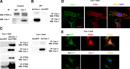
Figure 4.
PTEN associates with caveolin-1 in control fibroblasts, but an association cannot be detected in IPF fibroblasts. A: Caveolin-1 was immunoprecipitated from control and IPF fibroblasts, and Western analysis for PTEN or caveolin-1 was performed (data shown for control only). B: IPF fibroblasts were infected with an adenoviral vector containing wild-type caveolin-1 construct. Cells infected with empty vector (Ad-GFP) served as a control. Caveolin-1 was immunoprecipitated, and Western analysis for PTEN and caveolin-1 was performed. C: Caveolin-1–null fibroblasts overexpressing wild-type caveolin-1 and PTEN constructs together (left) or wild-type caveolin-1 alone (right) were seeded on polymerized collagen. Caveolin-1 or PTEN was immunoprecipitated and Western analysis for PTEN, and caveolin-1 was performed. D and E: Shown is confocal microscopic immunofluorescent analysis of caveolin-1 and PTEN expression in caveolin-1–null fibroblasts reconstituted with caveolin-1 (D) or empty vector (E; GFP control) and seeded on polymerized collagen. Arrows denote colocalization of PTEN and caveolin-1 at the membrane in caveolin-1–null cells reconstituted with caveolin-1. Scale bar = 20 μm. Data are representative of three independent experiments.

