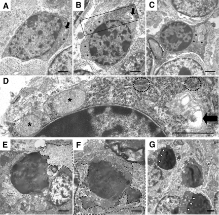
Figure 5.
Distinctive ultrastructural changes displayed by CGNs in Tg(ΔCR)/Prn-p+/0 mice. Electron microscopic images of CGNs in Tg(ΔCR)/Prn-p+/0 mice at P12 (A–D), P13 (E and F), and P20 (G). D shows a higher magnification of the area within the box in B. At P12, dying granule cells exhibit nuclear abnormalities such as matrix condensation and small, irregular dispersed chromatin clumps (A–D). Other cellular changes include cytoplasmic matrix condensation, swelling of mitochondria (asterisks), clustering of ribosomes (dashed circles), and dilation of the Golgi apparatus (black arrows). At P13, cells have continued to condense (E and F), with active phagocytosis occurring. Dashed lines outline phagocytic processes from adjacent glial cells. At P20, CGNs display similar morphological abnormalities. G: Whereas cell bodies become progressively shrunken, the nuclear membrane remains intact (white arrowheads). Scale bars in all panels: 1.1 μm.

