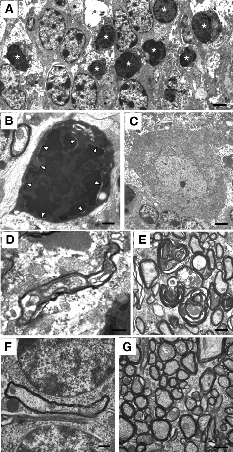
Figure 7.
PrPΔ32-134 induces nonapoptotic death of CGNs as well as axonal pathology. Ultrastructural analysis of the cerebella of clinically ill Tg(F35)/Prn-p0/0 mice at P32 (A and C–E) and P65 (B). A: Clusters of degenerating granule cells (asterisks) exhibit morphological characteristics similar to those seen in Tg(ΔCR) mice, including cellular shrinkage, darkening of the cytoplasmic matrix, and condensation of chromatin into interconnected clumps throughout the nucleus, with preservation of the nuclear membrane. B: Degenerating CGN displaying markedly condensed cytoplasm and accumulation of chromatin clumps but with maintenance of an intact nuclear membrane (arrowheads). C: Purkinje cell bodies remain normal. D: Swollen and dystrophic Purkinje cell axons in the granule cell layer contained vacuolated axoplasm. E: Axons in the cerebellar white matter display disintegrating myelin sheaths. Purkinje cell axons (F) and cerebellar white matter (G) of a P30 Prn-p0/0 control mouse are normal. Scale bars: 2.5 μm (A and C); 0.6 μm (B, D, and F); 1.1 μm (E and G).

