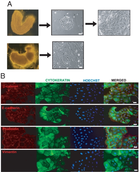
Figure 1.
Isolation of epithelial and stromal endometrial cells from mouse uterus. A, left: representative images of the epithelial sheet (top) and the rest of the uterine horn fragment (bottom) after digestion with trypsin and separation of two parts. Right, representative images of isolated epithelial and stromal cells cultured in 2D monolayer cells for 24 hours. B: Double immunofluorescence showing positive labeling of 2D monolayer epithelial cells with cytokeratin, E-cadherin, β-catenin, and phalloidin antibodies and negative staining for vimentin. In all immunofluorescence experiments cells were counterstained with Hoechst dye. White scale bar = 20 μm.

