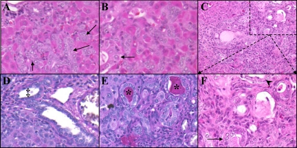
Figure 10.
Atypical ductular hyperplasia within Wnt1 shRNA–treated animals. A: H & E Day nine. B: H & E Day 13. C and F: H & E Day 21. D: Periodic acid-Schiff staining of 2AAF/PHx Day nine. E: Periodic acid-Schiff staining of Wnt1 shRNA–treated animal day 21. F: Magnification of outlined area of C allowing for closer examination of ADH. Treatment with shRNA to Wnt1 in the 2AAF/PHx model induces oval cells to undergo differentiation toward a ductular lineage. Ducts that remain retain a fairly normal cuboidal morphology (A and B, arrows) until 15 to 21 days post PHx. At this point, ADH ensues. As seen in D, some ducts undergo transformation into columnar (arrow) and even squamous (arrowhead) phenotypes. The atypical ducts are mucin positive (asterisks), whereas, ducts found in the standard 2AAF/PHx protocol are mucin negative (‡). This indicates the atypical ducts are no longer of a biliary lineage. Magnification in A, B, D, E, and F: ×40; in C: ×20.

