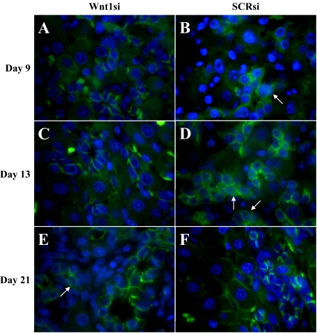
Figure 11.
β-catenin staining of shRNA-treated animals. A, C, and E: Wnt shRNA–treated animals; B, D, and F: SCR shRNA–treated animals; A and B: Day nine. C and D: Day 13. E and F: Day 21. β-catenin activation and nuclear translocation (arrows) is present in numerous oval cells at the peak of oval cell numbers (Day 9) in the 2AAF/PHx model (data not shown) and SCR shRNA–treated animals and are very prevalent during the initiation of oval cell differentiation (Day 13). Oval cells of Wnt1 shRNA–treated animals do not translocate β-catenin at the peak of oval cell numbers, but instead a few rare cells can be identified in areas of ADH on Day 21.

