Figure 6.
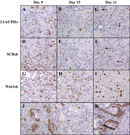
Ki-67 comparison of 2AAF/Phx versus shRNA-treated animals. A: 2AAF/PHx Day nine. B: 2AAF/PHx Day 15. C: 2AAF/PHx Day 21. D: SCR shRNA Day nine. E: SCR shRNA Day 15. F: SCR shRNA Day 21. G: Wnt1 shRNA Day nine. H: Wnt1 shRNA Day 15. I: Wnt1 shRNA Day 21. J: Wnt1 shRNA Day 21 area of ADH. K: Intestine (positive control); proliferation of oval cells nine days after PHx in shRNA-treated animals mimics that observed in 2AAF/PHx alone. In 2AAF/PHx alone and SCR shRNA–treated animals by day 15 proliferation has subsided as oval cells begin differentiating. Upon recovering from the influence of 2AAF, hepatocytes can be seen to divide 21 days after PHx in SCR and control animals (arrows). On the contrary, oval cells in Wnt1 shRNA–treated animals have an approximately 47% decrease in proliferation on Day nine but increase their proliferation rate by Day 13 (data not shown) and continue to proliferate 15 days after PHx (arrow heads). Also, in Wnt1 shRNA–treated animals hepatocytes that have begun to recover from the influence of 2AAF exhibit a very high proliferative rate 21 days after PHx (arrows), whereas under normal conditions the liver has completely recovered and division is unnecessary 21 days after PHx. It can also be observed that the cells within sites of atypical ductular hyperplasia are also rapidly dividing 21 days after PHx. Magnification, ×20.

