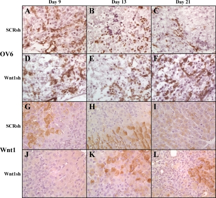
Figure 8.
OV6 and Wnt1 staining of the livers of shRNA-treated animals. A–F: OV6 staining of fresh frozen sections. G–L: Wnt1 staining of paraffin sections. A–C and G–I: Wnt1 shRNA–treated animals. D–F and J–L: SCR shRNA–treated animals; A, D, G, and J: Day nine. B, E, H, and K: Day 13. C, F, I, and L: Day 21. Oval cells from 2AAF/PHx and SCR shRNA–treated animals express OV6 in high levels early in oval cell induction before their differentiation, and pericentral hepatocytes express Wnt1 (2AAF/PHx data not shown). In vivo treatment of animals with Wnt1 shRNA on Days three and six post PHx, inhibits Wnt1 expression until at least Day 13. After 21 days, Wnt1 expression returns to interzonal and pericentral hepatocytes. OV6 levels stay fairly constant during Wnt1 shRNA treatment whereas in SCR shRNA–treated animals the numbers of OV6-positive cells diminish after 13 days, and the cells are practically nonexistent as of Day 21. SCR shRNA staining is indistinguishable from 2AAF/PHx staining (data not shown) Magnification, ×40.

