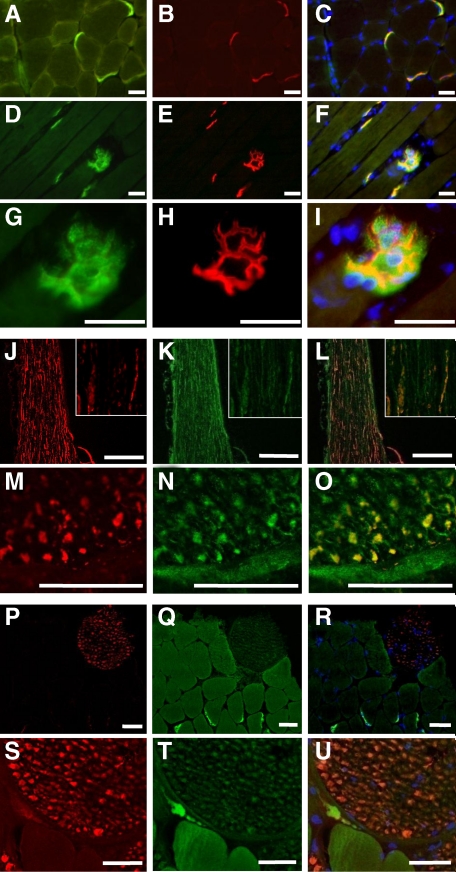
Figure 2.
DNAJB2 immunoreactivity in normal control C57BL/6 mice. A–I: Serial transverse (A–C) and longitudinal (D–I) sections of the tibial anterior muscle in wild-type mice. DNAJB2 is expressed at the NMJ, shown by double staining with DNAJB2 (green; A, D, G), α-bungarotoxin (red; B, E, H), and their co-staining (yellow; C, F, I). The branched structure of the NMJ postsynaptic membrane observed in (D–F) is shown at a larger magnification in (G–I). Nuclei are stained with 4,6-diamidino-2-phenylindole (DAPI; blue; C, F, I). Scale bars = 50 μm. J–O: Isolated sciatic nerve is shown on longitudinal (J–L) and transverse (M–O) sections, with anti-neurofilament (red; J, M), DNAJB2 (green; K, N), and double staining (orange-yellow; L, O). The inset represents a larger magnification of a part of the nerve shown in J–L. Bars indicate a magnification of 200 μm (J–L) and 25 μm (M–O). P–U: A small nerve branch on a transverse section through the psoas muscle reveals anti-DNAJB2 immunofluorescence (Q, T) that is weaker than in the NMJ. Axonal colocalization (orange; R, U) of anti-DNAJB2 (green; Q, T) with anti-neurofilament (red; P, S) is found. Scale bars = 50 μm.

