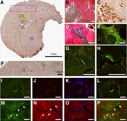
Figure 3.
Expression of DNAJB2 in dystrophic muscle of the mdx4Cv mice. A: DNAJB2 staining of transverse section of tibial anterior muscle in mdx mouse. The green box in (A) is seen at a higher magnification in (B–C) by H&E (B) and anti-DNAJB2 immunohistochemical stains (C). Similarly, the blue box in (A) is shown at a higher magnification in (D–E) by H&E stain (D) and DNAJB2 immunofluorescence (E). Notice that DNAJB2 is diffusely expressed in the sarcoplasm and at the sarcolemma of small regenerating fibers (blue arrowhead), whereas in slightly larger regenerating fibers its expression is mainly observed at the sarcolemma (red arrowhead). In mdx mouse, strong expression of DNAJB2 is revealed at the neuromuscular junction (green arrowhead in C). DNAJB2 in NMJs is seen further in the purple box (A) and at a higher magnification (F), and by confocal imaging (G–H; DNAJB2 in green. Nuclei are shown in red (propidium iodide)). I–L: Double immunostaining at NMJs is shown: anti-DNAJB2 in green (I), α-bungarotoxin in red (J), α-bungarotoxin merged with DAPI (blue, K) and merged with anti-DNAJB2 (yellow, L). M–P: Co-expression of DNAJB2 and α-bungarotoxin is seen (white arrows) at the sarcolemma of regenerating fibers (DNAJB2, green, M; α-bungarotoxin, red, N; α-bungarotoxin merged with DAPI, blue, O; α-bungarotoxin merged with anti-DNAJB2, yellow (P). Scale bars = 50 μm, except (H) in which the bar corresponds to 25 μm.

