Figure 4.
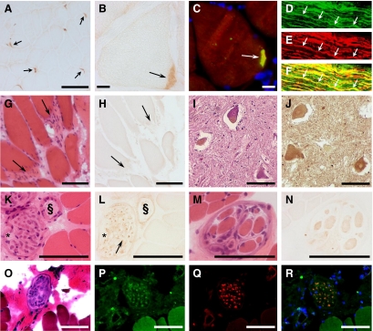
DNAJB2 immunoreactivity in normal human tissues. A–B: NMJs (arrows) in control muscle (control 9; see Table 1) show strong immunoreactivity for the DNAJB2 antibody, revealed by immunohistochemical (A–B) and immunofluorescence (C) techniques. Co-localization of DNAJB2 with α-bungarotoxin at the neuromuscular junction is shown in yellow (C arrow). D–F: A longitudinal section through a normal sural nerve (control 10) reveals axonal colocalization (yellow) (F) of DNAJB2 (D) and neurofilament (E). One axon is indicated by arrows. G–H: Myotendinous junctions (arrows) in a normal control biopsy (control 2) reveal no immunoreactivity for anti-DNAJB2, as indicated on serial transverse sections at H&E stains (G) and anti-DNAJB2 immunoreactivity (H). I–J: Transverse sections through the ventral horn of the spinal cord (control 12) at H&E (I) and DNAJB2 (J) stains, reveal anti-DNAJB2 immunoreactivity in the soma of motoneurons and in axons. K–L: Normal control skeletal muscle (control 6) shows muscle fibers with a very weak sarcoplasmic immunoreactivity using the anti-DNAJB2 antibody, at serial transverse sections with H&E (K) and DNAJB2 (L) stains. Reactivity for anti-DNAJB2 in the axons (arrow) of a transversally cut perimysial nerve (asterisk) and in the wall of a perimysial blood vessel (§) are shown. M–N: Intrafusal fibers of a muscle spindle are slightly stained with anti-DNAJB2, shown on transverse serial sections through normal control muscle (control 1) by H&E (M) and DNAJB2 (N) stains. O–R: Serial transverse muscle sections (control 6) show a perimysial nerve at H&E stains (O), and fluorescence staining with anti-DNAJB2 (P), anti-neurofilament (Q), and their co-staining (R). The myonuclei are stained in blue (DAPI). Scale bars = 100 μm, except (B, C) in which the bar corresponds to 10 μm.

