Figure 3.
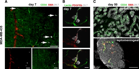
Blood vessel types involved in cancer cell extravasation and growth. Blood vessel types were analyzed by immunofluorescence. Representative images for MDA-MB-435 cells are shown. A: Day seven: in the brain parenchyma, cancer cells (white, arrows) arrest and extravasate exclusively from capillaries or postcapillary venules positive for CD34 and lacking smooth muscle cells (no smooth muscle actin (SMA) signal). Scale bars: 100 μm. B: Day seven: blood vessels from which cancer cells (gray) extravasate are surrounded by platelet-derived growth factor receptor β (PDGFRb)-positive pericytes. Top panel: BS-1 lectin; middle panel: anti-PDGFRb; and bottom panel: merge. Scale bar: 20 μm. C: Day 50: intraparenchymal metastases grow around co-opted capillaries lacking smooth muscle cells. Leptomeningeal metastases contain capillaries as well as larger, smooth muscle cell-positive vessels. Scale bars: 100 μm.

