Figure 5.
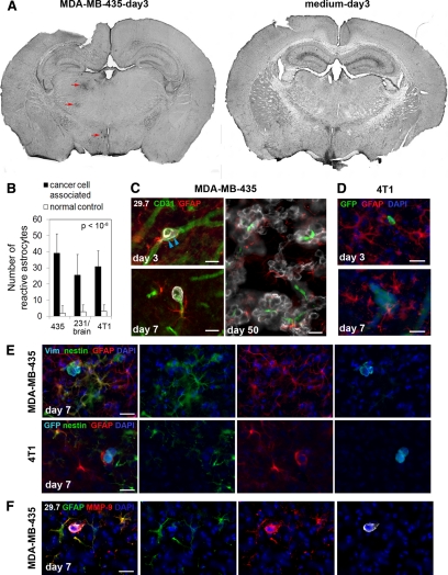
Cancer cell invasion induces strong astrocytic responses. Astrocytes were investigated by immunofluorescence staining. A: Left, On day three after cancer cell injection into the left carotid artery, GFAP in astrocytes is already up-regulated strongly in the vicinity of intravascular arrested cancer cells (MDA-MB-435, red arrows). Astrocyte activation can be detected in the left hemisphere in brain overview sections, whereas the corresponding area of the contralateral hemisphere is devoid of GFAP reactivity. Right, Also, no GFAP activity was found in the brain of control animals injected with medium alone. B: Number of reactive astrocytes three days after carotid artery injection of cancer cells, quantified within the 150-μm distance from cancer cells (cancer cell associated) and within the corresponding region of the contralateral hemisphere that lacks cancer cells (normal control). C: Activated astrocytes with thick processes and up-regulated expression of GFAP are detected next to MDA-MB-435 cancer cells that are still intravascular. Note the cytoplasmic protrusions of cancer cells on day three postinoculation that apparently cause stretching of the vessel wall (blue arrowheads) (day three, upper left panel). GFAP-positive astrocytes stay close to extravasated tumor cells (day seven, lower left panel). Reactive astrocytes persist close to cancer cells throughout their development into macrometastases (day 50, right panel). Scale bars: 20 μm. D: Activated astrocytes are also present in the vicinity of 4T1 breast cancer cells injected into the carotid artery of syngeneic BALB/c mice. Scale bars: 20 μm. E: In addition to GFAP up-regulation, some reactive astrocytes simultaneously express nestin. Merged images are shown on the left. Human vimentin or GFP (light blue), nestin (green), GFAP (red), and 4′,6′-diamidino-2-phenylindole (DAPI) (dark blue). Scale bar: 20 μm. F: Strong up-regulation of MMP-9 is detected in reactive astrocytes located in the immediate vicinity of extravasated MDA-MB-435 tumor cell. Scale bar: 20 μm.

