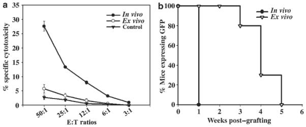
Figure 3. Analysis of GFP-specific CTL responses.

(a) Stimulated lymphocytes (effector cells) were harvested and viable effector cells were mixed with GFP-expressing syngeneic fibroblasts at different effector:target ratios (50:1 to 3:1) and incubated for 5 h. GFP-tolerant mice grafted with GFP+ keratinocyte were used as controls. Specific lysis of target cells was determined in triplicates using LZ-expressing syngeneic fibroblasts as control targets. (b) Mice were transduced in vivo (n = 4) with INV-GFP or ex vivo (n=6) with LZRS-GFP, and at 7 weeks posttransduction when GFP expression was lost in all mice, they were challenged with transplantation of GFP expressing keratinocytes. Mice were monitored weekly and GFP-expression in the grafted area was scored. A ≥90% decrease in surface GFP was taken to indicate graft loss.
