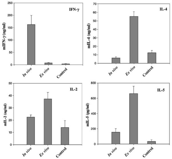
Figure 4. In vitro cytokine secretion by lymphocytes of transduced mice.

Splenocytes and lymphocytes from axillary lymph nodes were isolated at 2 or 5 weeks posttransduction from in vivo- or ex vivo-transduced mice, respectively. Controls are Gad-GFP mice transplanted with GFP+ keratinocytes. Lymphocytes were cultured with mitomycin-treated GFP-expressing splenocytes for 72 h. Cell-free supernatant was collected and analyzed by cytokine-specific ELISA as indicated in the graph (n = 6). Results are expressed as mean±SEM.
