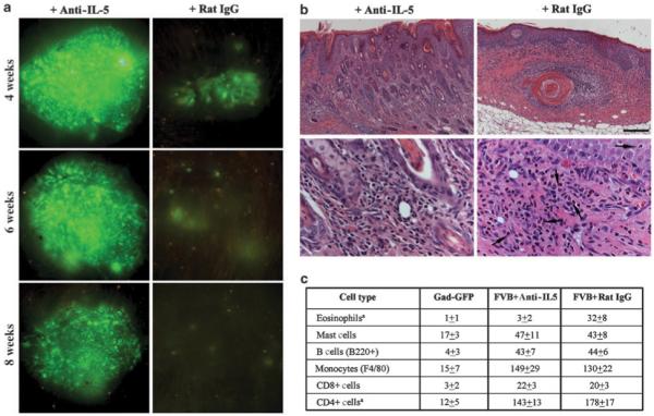
Figure 6. Effect of IL-5blockade on transgene expression in ex vivo-transduced mice.

(a) Prolonged surface GFP expression in a representative mouse receiving a single injection of 30 μg of anti-mIL5 (TRFK5) followed by grafting of ex vivo-transduced keratinocytes. In mice pretreated with 30 μg of control rat IgG, surface GFP is almost lost at 4 weeks postgrafting. (b) Histological analysis of transduced skin sections obtained at 4 weeks posttransduction and stained with hematoxylin/eosin staining shows suppression of eosinophil infiltration in mice treated with anti-IL5. Lower panels are at higher magnification to show eosinophilic infiltrates. Eosinophils (arrows) are easily identified by their plurilobed nuclei and the intense eosin staining of their cytoplasm. (c) Summary of distribution of inflammatory cells in skin reconstituted on GFP-tolerant mice or FVB mice pretreated with anti-IL-5 antibody or Rat-IgG. Cellular infiltrates were assessed in four randomly selected fields at × 400 original magnification and are expressed as mean±SEM.
