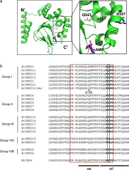
Fig. 5.
The location of R557 and Q543 in tertiary structure and amino acid sequence alignment. (a) Ribbon diagram of the cytoplasmic C-terminal region of AtCNGC11/12 (AtCNGC12) (left panel) and close-up of the indicated area of the left panel (right panel). R557 is located in the αC-helix and Q543 is located in the αB-helix of the CNBD. cAMP is indicated by pink colour. (b) Alignment of the area of R557 with 20 Arabidopsis CNGCs and tobacco NtCBP4. NCBI:AF079872, AtCNGC3:CAB40128, AtCNGC11:AAD20357, AtCNGC10:AAF73128, AtCNGC13:AAL27505, AtCNGC1:AAK43954, AtCNGC12:AAd23055, AtCNGC12 (Ws ecotype):EU541495, AtCNGC6:AAC63666, AtCNGC9:CAB79774, AtCNGC5:T52573, AtCNGC7:AAG12561, AtCNGC8:NP_173408, AtCNGC15:AAD29827, AtCNGC14:AAD23886, AtCNGC17:CAB81029, ATCNGC16:CAB41138, AtCNGC18:CAC01886, AtCNGC19:BAB02061, AtCNGC20:BAB02062, AtCNGC2:CAC01740, AtCNGC4:T52574. The black box indicates the position of R557. The red box indicates the CaM binding domain and bold characters highlight the critical four amino acids for the CaM binding suggested by Arazi et al. (2000). The location of Q543 (S136) is indicated by a black dot.

