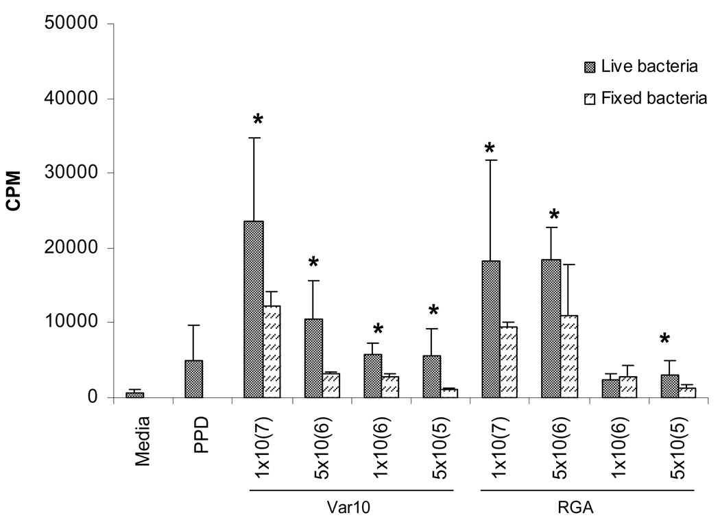
Figure 1.
Live Leptospira- induced proliferation of control PBMC. PBMC were cultured in microtiter wells at 2×10⁵/well with one of the following: 1) medium only, 2) PPD (5µg/mL), or 3) Leptospira, or 4) formalin- fixed Leptospira. Cell proliferation was assessed at 6 days by measuring the amount of [3H] thymidine incorporation. Results are from one experiment that was representative of 7 independent experiments (7 different controls). * P<0.05 live vs fixed bacteria.

