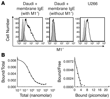
Figure 1. M1′-specific antibody specifically binds human M1′ in the context of membrane IgE with high affinity.
(A) M1′-specific antibody 47H4 bound human membrane IgE-transfected Daudi B cells but not Daudi transfectants expressing membrane IgE that lacks the M1′ sequence. M1′-specific antibody 47H4 also bound the U266 myeloma cell line, which naturally expresses low levels of membrane IgE. Isotype control antibody staining is shown as the gray areas, and 47H4 antibody staining is shown as black lines. (B) Scatchard analysis of M1′-specific antibody 47H4 binding to membrane IgE-transfected Daudi cells. The left panel shows a competition binding curve, where each data point represents the ratio of iodinated 47H4 antibody bound to cells/total iodinated and unlabeled 47H4 antibody on the y axis vs. the total concentration of iodinated and unlabeled 47H4 antibody on the x axis. The right panel shows a Scatchard plot, where each data point represents the ratio of iodinated 47H4 antibody bound to cells/unbound iodinated 47H4 antibody on the y axis vs. the concentration of bound iodinated 47H4 antibody on the x axis The mean binding affinity ± SD from 2 separate experiments is 0.54 ± 0.02 nM.

