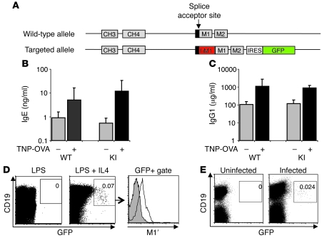
Figure 2. Human M1′/GFP knockin mouse has normal antibody responses and generates M1′+ GFP+ IgE B cells.
(A) Targeting scheme for insertion of human M1′ and a bicistronic GFP reporter gene in the mouse IgE locus. Human M1′ (red) is inserted into the mouse M1 exon splice acceptor site in-frame, with the M1 exon coding sequence. An IRES-GFP (green) bicistronic reporter gene is inserted 26-bases downstream of the end of the M2 exon. Wild-type and M1′ knockin (KI) mice immunized with TNP-OVA/alum have identical baseline and immunization-induced serum (B) IgE and (C) IgG1, as measured by ELISA. (D) Ex vivo culture of M1′ knockin mouse splenocytes with LPS and IL-4, but not LPS alone, induced M1′+ GFP+ B cells. Numbers indicate the percentage of CD19+ GFP+ cells and are representative of at least 3 experiments. Flow cytometry data from LPS+IL-4–stimulated cultures in the center panel was further gated on GFP vs. CD19 expression as defined by the box and analyzed for M1′ expression as indicated by the arrow to the right-hand panel. Isotype control antibody staining is shown as the gray areas, and 47H4 antibody staining for M1′ expression is shown as black lines. (E) A small population of GFP+ B cells was detectable in the mesenteric lymph nodes of N. brasiliensis–infected M1′ knockin mice. Numbers indicate the percentage of CD19+ GFP+ cells and are representative of at least 3 experiments.

