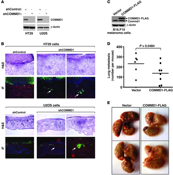
Figure 2. COMMD1 repression promotes tumor invasion.
(A–C) HT29 or U2OS cells were infected with lentiviruses expressing short hairpin sequences targeting COMMD1 (shCOMMD1) or a control gene (shControl). (A) Western blot analysis demonstrating the decrease in COMMD1 levels in the cell lines used is shown. These cells were tested for invasion of the chick CAM, as described in the Methods section. (B) Representative images of H&E and immunofluorescence staining (IF) are shown (laminin, red; DAPI, blue; cancer cells, green). Cell invasion into the CAM is indicated with white arrows (original magnification, ×200 [HT29]; ×100 [U2OS]). (C–E) B16.F10 mouse melanoma cells were stably transfected to express COMMD1-FLAG (as shown in the Western blot in C). These cells were subsequently injected into the tail vein of syngeneic C57BL6 mice (2 × 10⁵ cells/mouse), and (D) after 15 days, the mice were sacrificed, and the number of lung metastasis per mouse was counted (the horizontal bars represent the mean in each group). The Student’s t test was used for statistical analysis. (E) Representative images of the lungs of these mice are shown.

